Richard Cooper

Aug 012009
 

J. Appl. Cryst. (2009), 42, 798-809.    [ doi:10.1107/S0021889809024376 ]

A new method is presented for handling errors on crystallographic data. In single-crystal diffraction experiments, two variance-covariance matrices are present, one for the cell parameters and the second for the refined parameters (atomic coordinates and anisotropic displacement parameters). These two matrices can be combined so that errors on derived parameters, such as bond distances, bond angles and TLS tensors, may be calculated more simply. The new method works for all space groups but there are limitations on its application to triclinic space groups. The method allows errors to be transformed between space groups.

Publisher copy: IUCr

May 072009
 

Tet. Asym. (2009), 20, 712-717.    [ doi:10.1016/j.tetasy.2009.02.025 ]

Advances in hardware and software have made X-ray crystallography even more attractive as the first-option method for structure analysis. For most organic materials containing up to 100 non-hydrogen atoms, getting from the initial visual examination of the sample to producing publication-ready tables and pictures should usually be achievable in a single morning. Improvements in hardware have also increased reliability of the determination of absolute configuration. A recently published new algorithm may extend the range of applicability of the method.

Le Coup de Roi

Le Coup de Roi: making two chiral pieces from an achiral whole

Feb 172009
 

Bob graduated from St Edmund Hall (Oxford) having completed his Part II year doing copper chemistry with Francis Rossotti. He worked for his D. Phil. (still on copper chemistry) with Keith Prout and Francis, and it was during this work that he became interested in crystallography.

For one of the materials he worked on, aquo(maleato)copper(II), he observed “When the diffraction pattern was indexed, it became apparent that the crystals were unlikely to be orthorhombic, as a strange set of absences were found”. The crystals were twinned. “overlapped reflections were arbitrarily assigned half the measured intensity until a program was written which would include both components in the least squares”. “as there was not sufficient space [memory] it was necessary to rewrite the [AUTOCODE] program in [English Electric Leo Marconi] KDF9 machine language”. AUTOCODE was a symbolic language, rather like a simplified FORTAN. Machine languages are basic to the electronics of the computer, and the programmer has the power and the responsibility of working hands-on with every memory location, even to the extent of synchronising the calculations with the revolutions of the bulk storage devices. So began Bob’s life with computers, and his exceedingly productive partnership with John Rollett.

Immediately after writing his DPhil thesis in 1969, Bob was awarded a fellowship from the Accademia Nazionale dei Lincei in collaboration with the Royal Society which enabled him to work in Rome. There, he set about working with Riccardo Spagna re- implementing the Rollett AUTOCODES in FORTRAN. This program included features such as “riding” and rigid-body constraints, and some of the underlying data-structure can still be found in CAOS (Cerrini S. & Spagna R. (1977) Crystallographic software for a mincomputer, IV Eur. Crystallgr.Meet., Oxford, UK, Abstract A- 212).

On Bob’s return to Oxford he worked with Rollet and Prout, again re-writing the program from scratch but building upon his experiences in Rome. This new program, called CRYSTALS, could handle up to 9 twin components and had a good range of restraints (including facilities now often called SIMU and DELU). Perhaps the most novel feature was “user-defined restraints”, in which the user could define their own equation of restraint as part of the input data, which was then analytically differentiated by CRYSTALS. The equation parser and differentiating engine were all written in beautiful FORTRAN, and are still working, largely unmodified, in the current version of CRYSTALS. Bob’s attitude to programming combined a meticulous attention to detail with a far reaching ability to plan on an expansive scale.

After his Post Doc, Bob started work for Oxford University Computing Service, writing software for data-archiving. However, he continued to work on CRYSTALS whenever he could, and completely re-wrote the underlying data management for a third time when the university upgrade its mainframe to an International Computers Limited (ICL) 2980.

In about 1979 Bob left Oxford to work for Control Data Corporation, implementing meteorology programs on their supercomputers. Apart for a brief period in the 1980’s when he worked with Keith Davies at Chemical Design, Bob has spent most of his career implementing very large FORTRAN program systems, and in recent years modernising massive legacy packages. Weather forecasting may have profited from his work, but there is no doubt that crystallography lost an outstanding programmer when Bob left Oxford.

When not working with computers, Bob was a dependable drinking companion and a formidable Bar Billiards enthusiast. Some of us still remember Bob and George Sheldrick working with other young crystallographers to try to drink the bar dry at ECM 4 in Oxford in 1977. His brilliance as a scientist did not spoil his personality – he as always modest, amiable and good fun.
Apr 082008
 

J. Appl. Cryst. (2008), 41, 531-536.    [ doi:10.1107/S0021889808005463 ]

Librational motion within a crystal structure distorts the measured bond distances and angles from their physical values. TLS analysis of a rigid molecule or a rigid part of a molecule allows the calculation of bond-length and angle corrections. Until now, no estimate of the error on these corrections has been available. A method is presented for propagating the errors on the anisotropic displacement parameters (ADPs) to the bond-length and angle corrections which are a function of the libration tensor. The numerical significance of approximations made during the calculation is discussed.

Publisher copy: IUCr

Apr 092007
 

Acta. Cryst. (2007), 63, 303-308. [ doi:10.1107/S0108768106055212 ]

A new polymorph of 2,4-dihydroxybenzoic acid is reported. The structure was characterized by multiple-temperature X-ray diffraction and solid-state DFT computations. The material shows a geometric pattern of hydrogen bonding consistent with cooperativity between the intermolecular carboxylic acid dimer and intramolecular hydrogen bonds. The presence of proton disorder within this hydrogen-bond system, which would support such a cooperative model, was not fully ruled out by the initial X-ray studies. However, solid-state calculations on the three possible end-point tautomers indicate that the dominant crystallographically observed configuration is substantially lower in energy than the other tautomers (by at least 9 kJ mol-1), indicating that no disorder should be expected. It is therefore concluded that no disorder is observed either in the intra- or intermolecular hydrogen bonds of the title compound and that the cooperativity between the hydrogen bonds is not present within the temperature range studied.

Electronic reprints

Publisher’s copy

Jan 012006
 

Francesco obtained his PhD in Rome and since 2004 is Assistant Professor at the Chemical Division of the Department of Pharmaceutical Science of the University of Catania, Italy. He spent some months during 2005 and 2006 in our lab to improve his knowledge of crystallography. His principal area of research is the comparison of experimental X-ray diffraction data with computationally simulated data. The prediction of crystal morphology, polymorphism, atomic displacement parameters (adp), as well as the physico-chemical properties of biologically active compounds are among his interests.

Oct 222004
 
Andrew Cowley demonstrates the Nonius Kccd diffractometer to the Science Club. There is a second diffractometer behind the group.

Figure 1: Andrew Cowley demonstrates the Nonius Kccd diffractometer to the Science Club. There is a second diffractometer behind the group

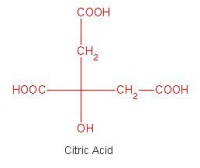
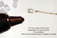
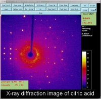
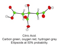
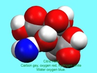
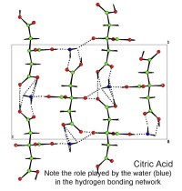
In October, Lynn Nickerson (Science Club Coordinator at Didcot Girls School) arranged for a small group to visit Chemical Crystallography in Oxford University’s new Chemistry Research Laboratory (Figure 1). The group was invited to bring some samples of common crystalline materials with them. The samples brought included cane sugar and citric acid (Figure 2). The girls used a polarising microscope to examine the crystals, and in the end selected an excellent crystal of citric acid for X-ray crystal structure determination. The crystal measured about 0.2 x 0.2 x 0.2 mm, and had to be ‘picked up’ on a fine nylon filament loop using a film of perfluoropolyether to hold it in place (Figure 3). The sample was put onto the Nonius kCCD automated diffractometer, cooled to -120°C and an X-ray diffraction image recorded (Figure 4). Dr Andrew Cowley collected a full data set in 40 minutes, which was processed by the Oxford crystallographic software CRYSTALS to reveal the structure of the acid (Figure 5 & 6). The hydrogen bonding network which holds the crystal together includes water of crystallisation, and is shown in Figure 7.


The molecular strcuture of citric acid

Figure 2: The molecular strcuture of citric acid

A single crystal of citric acid supported on a nylon loop. The ball point pen shows the scale

Figure 3: A single crystal of citric acid supported on a nylon loop. The ball point pen shows the scale

An X-ray diffraction image of citric acid The bright spots are Bragg reflections.

Figure 4: An X-ray diffraction image of citric acid The bright spots are Bragg reflections.


A single molecule of citric acid

Figure 5: A single molecule of citric acid

A 'space filling' image of citric acid. The blue atom is the oxygen atom of the water molecule which makes up part of the structure

Figure 6: A space filling image of citric acid. The blue atom is the oxygen atom of the water molecule which makes up part of the structure

A packing diagram of citric acid. The dotted lines are the hydrogen bond net work. These weak bonds help hold the crystal together

Figure 7: A packing diagram of citric acid. The dotted lines are the hydrogen bond net work. These weak bonds help hold the crystal together

Oct 132004
 

J. Chem. Inf. Comput. Sci. (2004), 44, 2133-2144. [ doi:10.1021/ci049780b ]

The crystallographically determined bond length, valence angle, and torsion angle information in the Cambridge Structural Database (CSD) has been made accessible by development of a new program (Mogul) for automated retrieval of molecular geometry data from the CSD. The program uses a system of keys to encode the chemical environments of fragments (bonds, valence angles, and acyclic torsions) from CSD structures. Fragments with identical keys are deemed to be chemically identical and are grouped together, and the distribution of the appropriate geometrical parameter (bond length, valence angle, or torsion angle) is computed and stored. Validation experiments indicate that, with rare exceptions, search results afford precise and unbiased estimates of molecular geometrical preferences. Such estimates may be used, for example, to validate the geometries of libraries of modeled molecules or of newly determined crystal structures or to assist structure solution from low-resolution (e.g. powder diffraction) X-ray data.

Electronic reprints

Publisher’s copy