Jun 152010
 

Dr David Watkin, an Honorary Member of the BCA, has been awarded the 2010 Fankuchen Award by the American Crystallographic Association. The award recognizes contributions to crystallographic research by one who is known to be an effective teacher of crystallography.

The Award will be presented at the ACA Annual Meeting in New Orleans 2011 at which David will also be delivering a Plenary Lecture.

Apr 152010
 

Cryst. Rev. (2010), 16(2), 133-144.    [ doi:10.1080/08893110903483238 ]

Recent developments in crystallographic error analysis are described. This report provides an introduction to more-formal work, originally published in Ref. (Haestier, J., J. Appl. Cryst. 2009, 42, 798). Prior to the main discussion, a brief overview of the normal distribution and error-propagation is provided, with some simplified examples to demonstrate the effects of covariance terms on error calculations. A new method is described for absorbing the cell-parameter errors into the variance-covariance matrix of the refined parameters. Problems occur for monoclinic and triclinic cell settings as the crystallographer must ‘choose’ how errors on the cell angles affect the coordinates. The choice has no effect on error-calculations on the internal geometry (bond-lengths, angles and torsion angles) of the structure, but may introduce a bias if the errors are used as weights.

Apr 012009
 
David Watkin gives the Lonsdale Lecture

David Watkin gives the Lonsdale Lecture

Every two years, the Young Crystallographers Group of the British Crystallographic Association nominate a speaker to give the prestigious Kathleen Lonsdale Lecture. Traditionally, they invite well respected scientist who has a good rapport with students. This year the Lonsdale Lecturer was David Watkin who is well known within the community, principally as a result of his involvement with the highly respected refinement software CRYSTALS, developed in Oxford and through the BCA biannual teaching school which he co-founded twenty-five years ago.

May 152008
 

J. Appl. Cryst. (2008), 41, 491-522.    [ doi:10.1107/S0021889808007279 ]

Most modern small-molecule refinement programs are based on similar algorithms. Details of these methods are scattered through the literature, sometimes in books that are no longer in print and usually in mathematical detail that makes them unattractive to nonprogrammers. This paper aims to discuss these well established algorithms in nonmathematical language, with the intention of enabling crystallographers to use their favourite programs effectively.

Oct 222004
 
Andrew Cowley demonstrates the Nonius Kccd diffractometer to the Science Club. There is a second diffractometer behind the group.

Figure 1: Andrew Cowley demonstrates the Nonius Kccd diffractometer to the Science Club. There is a second diffractometer behind the group

In October, Lynn Nickerson (Science Club Coordinator at Didcot Girls School) arranged for a small group to visit Chemical Crystallography in Oxford University’s new Chemistry Research Laboratory (Figure 1). The group was invited to bring some samples of common crystalline materials with them. The samples brought included cane sugar and citric acid (Figure 2). The girls used a polarising microscope to examine the crystals, and in the end selected an excellent crystal of citric acid for X-ray crystal structure determination. The crystal measured about 0.2 x 0.2 x 0.2 mm, and had to be ‘picked up’ on a fine nylon filament loop using a film of perfluoropolyether to hold it in place (Figure 3). The sample was put onto the Nonius kCCD automated diffractometer, cooled to -120°C and an X-ray diffraction image recorded (Figure 4). Dr Andrew Cowley collected a full data set in 40 minutes, which was processed by the Oxford crystallographic software CRYSTALS to reveal the structure of the acid (Figure 5 & 6). The hydrogen bonding network which holds the crystal together includes water of crystallisation, and is shown in Figure 7.
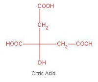
 

The molecular strcuture of citric acid
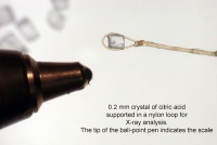
Figure 2: The molecular strcuture of citric acid

A single crystal of citric acid supported on a nylon loop. The ball point pen shows the scale

Figure 3: A single crystal of citric acid supported on a nylon loop. The ball point pen shows the scale
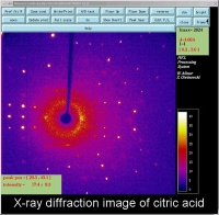
An X-ray diffraction image of citric acid The bright spots are Bragg reflections.
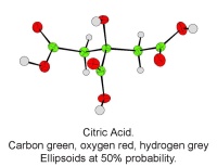
Figure 4: An X-ray diffraction image of citric acid The bright spots are Bragg reflections.


A single molecule of citric acid

Figure 5: A single molecule of citric acid
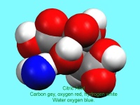
A 'space filling' image of citric acid. The blue atom is the oxygen atom of the water molecule which makes up part of the structure
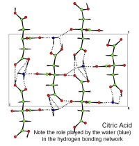
Figure 6: A space filling image of citric acid. The blue atom is the oxygen atom of the water molecule which makes up part of the structure

A packing diagram of citric acid. The dotted lines are the hydrogen bond net work. These weak bonds help hold the crystal together
Figure 7: A packing diagram of citric acid. The dotted lines are the hydrogen bond net work. These weak bonds help hold the crystal together