Mar 142014
 

2logos_frame_noletteringA-level students from a range of schools attended a one-day course at the Museum of the History of Science and the Department of Chemistry in Oxford to find out about the science and applications of crystallography. In the morning they discovered how symmetry plays important role in the structure and diffraction of crystals in a lecture by Prof. Brian Sutton of King’s College, London. Prof. Richard Cooper then gave an rapid overview of the history of the applications of crystallography from Pasteur’s discovery of chirality in the pre- X-ray diffraction world to Hodgkin’s determination of the structure of penicillin. Prof. Elspeth Garman took the students through the ups and downs of crystallographic research in the decades long attempt to grow one crystal of a virus protein in an attempt to fight the tuberculosis virus.

Split into groups, the students then visited the Department of Chemistry where they visited three different activities:

  • Rapid collection of diffraction data (under 10 minutes) and solution of the structure of fructose crystals with Dr. Amber Thompson in the X-ray facility.
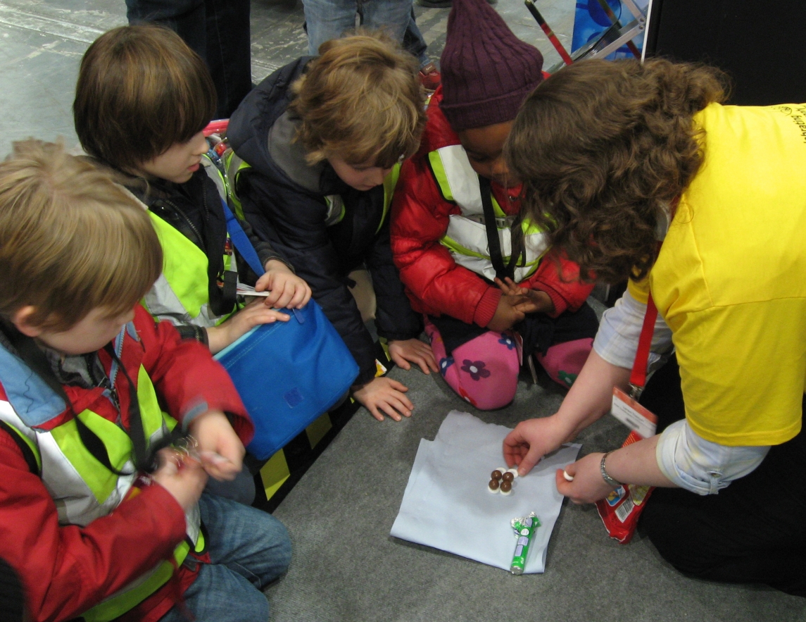
  •  Tasting how different crystalline structures (polymorphs) of cocoa butter in chocolate affect its texture and physical properties with Ms. Rachel Knight from Dirk Aarts’ research group (honorable mention to the one student who resisted temptation – having given up chocolate for lent!).
  • Exploring stereoisomers and enantiomers using physical models (including Pasteur’s tartrate ion) and discovering why mirror images of a molecule can have quite different smells.

Meanwhile back in the museum students visited the solar fuels outreach stand where they saw how crystallography can reveal the structures that nature uses to carry out photosynthesis, and, under the careful supervision of Johnny Brooks-Bartlett and Katharina Jungnickel from Biochemistry, they were able to carry out a recrystallisation of the protein lysozyme and watch while it grew in just a few minutes on a microscope slide.

May 232013
 

botanicThe University of Oxford Botanic Garden has organised a series of guided science walks through the Botanic Garden this summer.

Richard Cooper will be leading the first walk on 30th May entitled “How Do we Know what Molecules Look Like?”  A rich variety of molecules occur naturally in plants and some have incredible properties: from bitter tastes to anti-cancer activity. This walk will take in plants in the Botanic Garden from which interesting molecules have been discovered and describe how we can use analytical techniques to understand their shape and functions.

These guided walks will take place at 6.30pm at the Botanic Garden and will last approximately an hour followed by a glass of wine or soft drink. Tickets are avaliable from the University web site.

Mar 122013
 

The Big Bang Fair is a free educational event open to visiting school groups that happens in March every year moving round the country. It works with partner organisations across business and industry, government and academia to try and give a flavour of the real scale of engineering and science in the UK, aimed at showing young people (primarily aged 7-19) just how many exciting and rewarding opportunities there are out there for them with the right experience and qualifications.

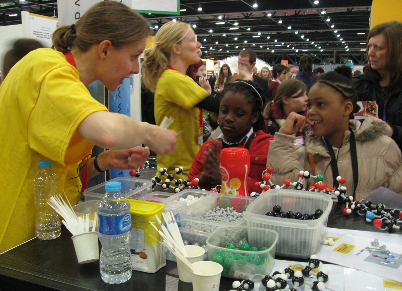
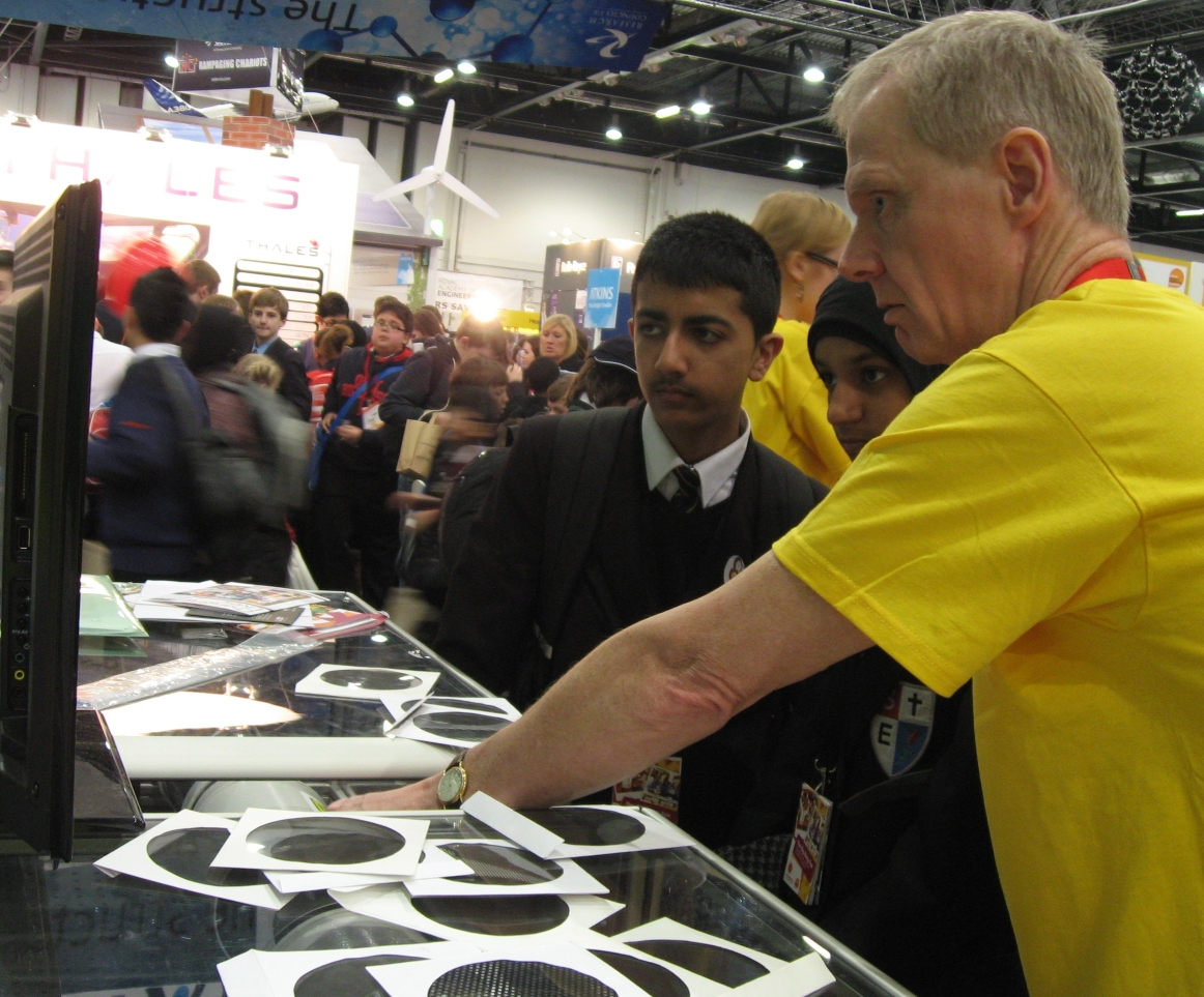
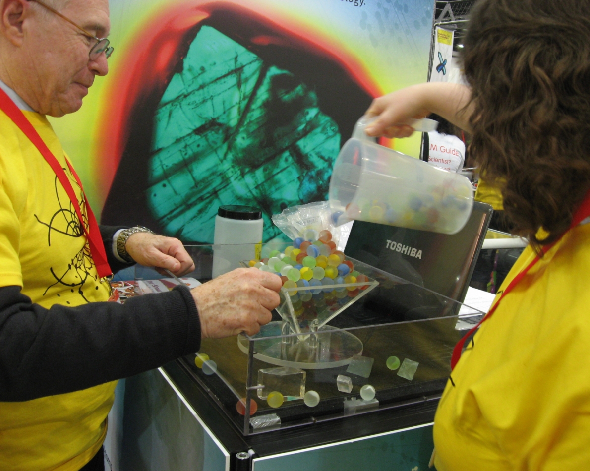
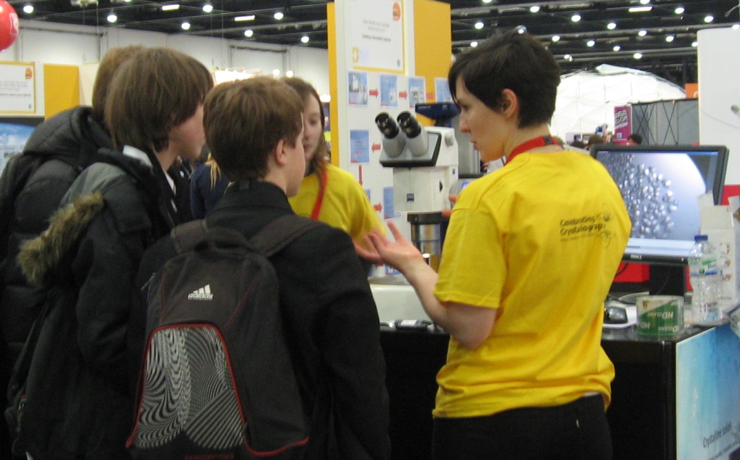
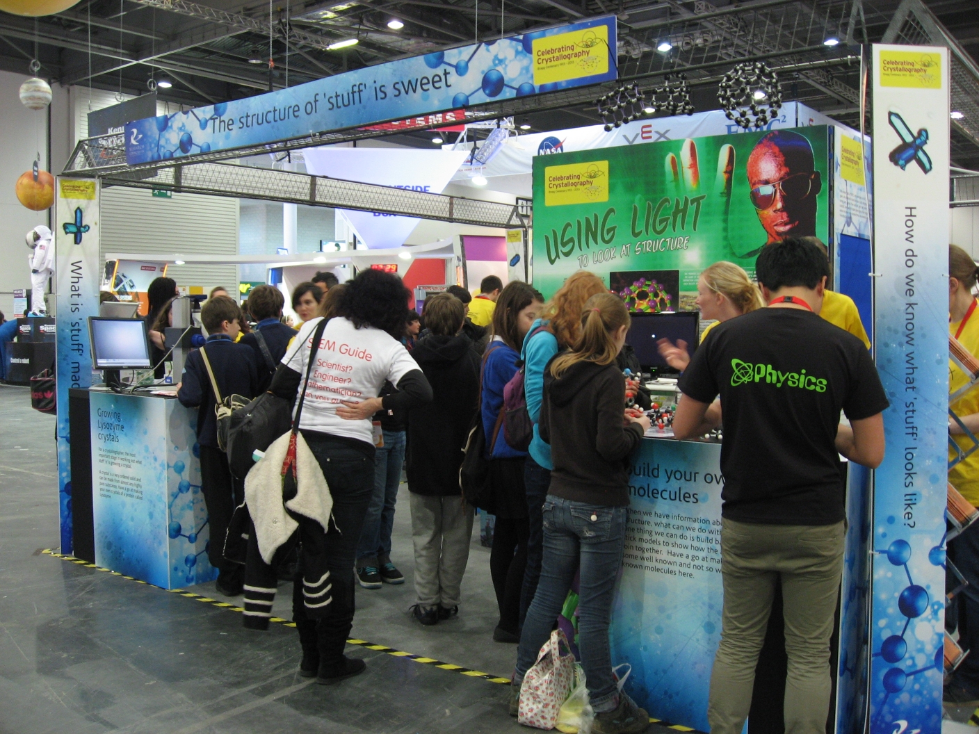
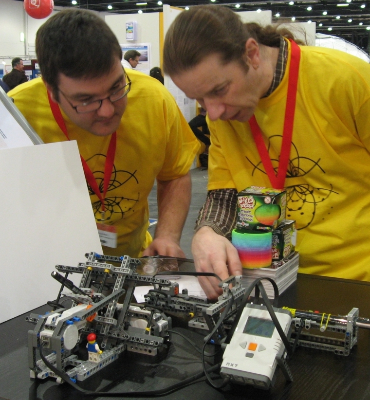
This year the Big Bang Fair is being held in the London, ExCeL Arena, 14th-17th March. Since 2013 is the Bragg centenary, STFC have very kindly funded a stand at this year’s fair, which will be totally dedicated to crystallography. The BCA, Diamond Light Source, ISIS and STFC have worked together to develop the stand designed to tell everyone how great crystallography is through the medium of hands on activities, lasers, and sweets. The fair is expecting 75,000 people (mostly children) through the doors over the course of four days, so Andrew Cairns, Josh Hill, Nick Funnell, Mike Glazer, George Pidgeon, Karim Sutton and Amber Thompson are all going along from Oxford to help out. Here are some photos of the first day.

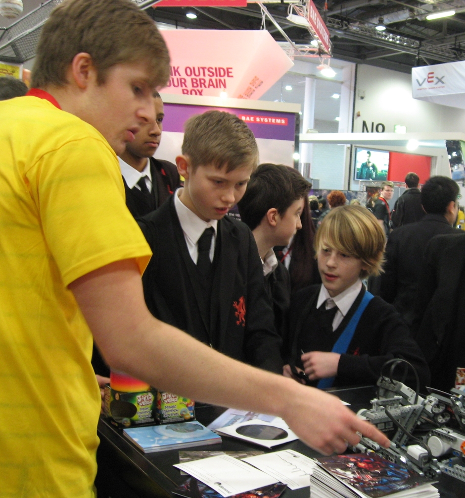
Lego Beamline

Two crystallographers check the interlocks on the Lego Beamline

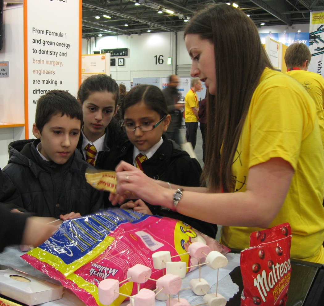
You are never too young to learn about packing...

You are never too young to learn about packing…

...especially when there's sweets involved!

…especially when there’s sweets involved!

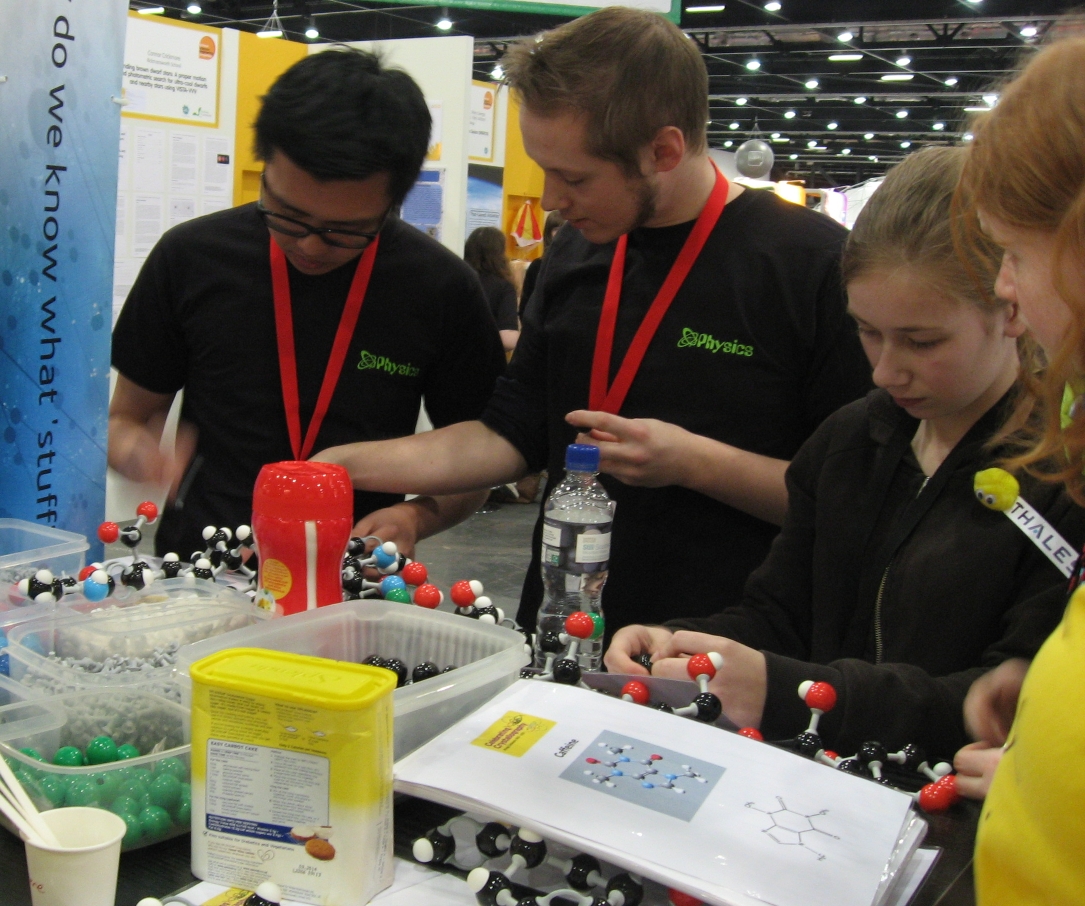
Teaching physicists chemistry

Teaching physicists chemistry

George demonstrates the Lego Beamline

George demonstrates the Lego Beamline

Smelly molecules

Smelly molecules

Demonstrating Fourier transforms takes concentration

Demonstrating Fourier transforms takes concentration

Growing a crystal, one marble at a time

Growing a crystal, one marble at a time

Protein crystals ar

Protein crystals are beautiful

The Crystallography stand at the Big Bang

The Crystallography stand at the Big Bang


The Big Bang Logo

Nov 292012
 

As part of the Bragg Centenary Celebrations, Melvyn Bragg and his guests discussed the history of crystallography, the study of crystals and their structure on “In Our Time” on BBC Radio 4. His guests were Prof. Judith A. K. Howard (University of Durham), Dr. Christopher Hammond (University of Leeds) and Prof. Mike Glazer (University of Oxford).

The program began with the work of Johannes Kepler in the 17th century, and focussed on the work of the father-and-son team the Braggs in 1912.  It also covered aspects of the the work of the German physicist Max von Laue who had proved that X-rays are a form of light waves and that it was possible to scatter these rays using a crystal and some of the most significant scientific findings of the last century – such as revealing the structure of DNA.

The program is available on line from the “In Our Time” website.

Nov 242012
 

Every year, Oxford Inspires organises an evening of social and cultural events in the centre of Oxford following the theme of Christmas Lights.  A lot of the institutions within the city get involved, and this year that included Amber, who was invited to give a presentation at the Snowflake exhibition held at the History of Science Museum.  The event was held on Friday, 23 November and the Museum opened at 6pm with talks from 7pm in the Gallery (that’s the basement to anyone else, so Amber felt at home).   Activities included:

  • Mirror Snowflakes – Create an infinite variety of symmetrical snowflakes with the help of a pair of mirrors.
  • Paper Crystals – Make your own snowflake decorations for Christmas.
  • Dr. Judith V. Field (Birkbeck College, London), “When Stars of Snow Fell on Kepler’s Coat
  • Dr. Amber L. Thompson (University of Oxford), “Why Is Snow So Beautiful?

The evening was very well attended with a very mixed audience.

Why is Snow So Beautiful?

Mar 302011
 

Rowena ScottIn-situ Diamond RingNot content with showing off the eye-catching brilliance of her new diamond engagement ring, final year Inorganic Chemistry student Rowena Scott wanted to reveal the beautiful symmetry of its atomic structure. She recorded an X-ray diffraction pattern from the diamond using one of the Chemistry Department’s new state of the art, SuperNova dual source X‑ray diffractometers.

Rowena’s current research project with Dr Simon Clarke involves the synthesis of iron chalcogenide superconductors. The structures of these materials are then determined using single crystal X-ray diffraction techniques.

Chemical Crystallography Service manager, Dr Amber Thompson said “We see a wide range of materials of varying crystallinity from many research groups in the Department of Chemistry, usually grown at the bottom of glass schlenks or flasks; it’s not often that such a lovely crystal turns up mounted in a band of gold”.

Diffraction pattern of the diamondThe diffracted X-rays were measured while rotating the diamond in an X-ray beam. The resulting diffraction pattern shows the position and intensity of the beams diffracted by the organised structure of carbon atoms in the crystal. The white rings overlaid on the image indicate the angles at which diamond peaks are expected to occur, which agree with the collected data.

Diamond diffracts X-rays so efficiently that the X-ray generator had to be run at 10% of its usual power in order to avoid overloading the X-ray detector.

Oct 222004
 
Andrew Cowley demonstrates the Nonius Kccd diffractometer to the Science Club. There is a second diffractometer behind the group.

Figure 1: Andrew Cowley demonstrates the Nonius Kccd diffractometer to the Science Club. There is a second diffractometer behind the group

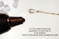
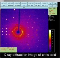
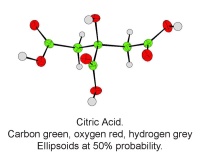
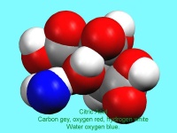
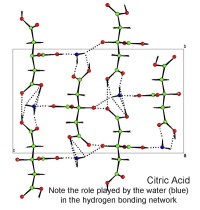
In October, Lynn Nickerson (Science Club Coordinator at Didcot Girls School) arranged for a small group to visit Chemical Crystallography in Oxford University’s new Chemistry Research Laboratory (Figure 1). The group was invited to bring some samples of common crystalline materials with them. The samples brought included cane sugar and citric acid (Figure 2). The girls used a polarising microscope to examine the crystals, and in the end selected an excellent crystal of citric acid for X-ray crystal structure determination. The crystal measured about 0.2 x 0.2 x 0.2 mm, and had to be ‘picked up’ on a fine nylon filament loop using a film of perfluoropolyether to hold it in place (Figure 3). The sample was put onto the Nonius kCCD automated diffractometer, cooled to -120°C and an X-ray diffraction image recorded (Figure 4). Dr Andrew Cowley collected a full data set in 40 minutes, which was processed by the Oxford crystallographic software CRYSTALS to reveal the structure of the acid (Figure 5 & 6). The hydrogen bonding network which holds the crystal together includes water of crystallisation, and is shown in Figure 7.


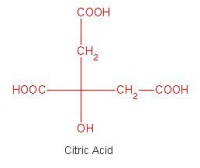
The molecular strcuture of citric acid

Figure 2: The molecular strcuture of citric acid

A single crystal of citric acid supported on a nylon loop. The ball point pen shows the scale

Figure 3: A single crystal of citric acid supported on a nylon loop. The ball point pen shows the scale

An X-ray diffraction image of citric acid The bright spots are Bragg reflections.

Figure 4: An X-ray diffraction image of citric acid The bright spots are Bragg reflections.


A single molecule of citric acid

Figure 5: A single molecule of citric acid

A 'space filling' image of citric acid. The blue atom is the oxygen atom of the water molecule which makes up part of the structure

Figure 6: A space filling image of citric acid. The blue atom is the oxygen atom of the water molecule which makes up part of the structure

A packing diagram of citric acid. The dotted lines are the hydrogen bond net work. These weak bonds help hold the crystal together

Figure 7: A packing diagram of citric acid. The dotted lines are the hydrogen bond net work. These weak bonds help hold the crystal together